A 53-year-old woman presents with worsening vascular Skin Lesions, fever, and a very high LDH. What is the underlying diagnosis? Explore this challenging case.

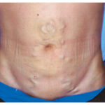
A 53-year-old woman presented with a 3-month history of worsening vascular skin lesions and a 1-month history of fever. On physical examination, diffuse telangiectasis, hyperpigmented plaques, and several ulcerated nodules (arrows) were observed on the skin across the chest and abdomen (left) and the legs. No palpable lymphadenopathy or hepatosplenomegaly was noted. Laboratory studies were notable for a lactate dehydrogenase level of 35664 U per liter (reference range, 120 to 250). A deep skin biopsy specimen from the abdomen showed intravascular aggregation of round, atypical lymphocytes (right, hematoxylin and eosin staining). Subsequent immunohistochemical staining was positive for CD20, PAX-5, and MUM-1 in the neoplastic cells. Which of the following is the most likely diagnosis?
What is the most likely diagnosis?
B-cell lymphoma, Dermatology, Intravascular lymphoma, Lactate Dehydrogenase, Oncology