A 26-year-old man presents with acute leg paralysis. Discover how imaging revealed a rare cardiac source and led to an emergency diagnosis.

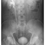
A 26-year-old man presented with sudden onset of severe pain in the legs and inability to move the left leg. On physical examination, he had complete loss of motor function in the left leg. Bedside ultrasonographic examination with color Doppler showed no blood flow in the distal aorta. Computed tomographic angiography of the abdomen revealed a saddle embolus at the aortoiliac junction (left). Emergency aortoiliac embolectomy was performed, and a gelatinous mass was removed. A subsequent transthoracic echocardiogram identified a heterogeneous mass in the left atrium (middle). On hospital day 2, cardiothoracic surgery was performed to remove the left atrial mass, and a villous, friable lesion was excised (right). Histopathology of the cardiac mass showed abundant mucopolysaccharide matrix with scattered nests of lepidic cells. What is the diagnosis?