Explore the case of an 82-year-old man with progressive weakness, weight loss, and bilateral adrenal masses, leading to an unexpected fungal diagnosis.

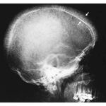
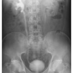
An 82-year-old man presented to the emergency department with a 3-year history of progressive generalized weakness. Four months before presentation, a left adrenal mass had been identified incidentally on computed tomography (CT) that had been performed to evaluate an episode of chest pain. During the month preceding presentation, he had lost 8 kg of weight and had become unable to sit up in bed. Physical examination was unremarkable. Laboratory studies were notable for a white-cell count of 2700 per cubic millimeter (reference range, 3700 to 10,500), a normal adrenal axis, and negative testing for HIV. Owing to concern for cancer, positron-emission tomography–CT of the whole body was performed and showed an adrenal mass on each side with fluorodeoxyglucose (FDG) uptake (left image shows a coronal view), a 17-cm-long spleen without FDG uptake (asterisk in left image), and no other abnormal findings. Subsequent biopsy of the left adrenal mass showed necrotizing granulomatous inflammation with intracellular fungal organisms (middle, arrows; hematoxylin and eosin staining) that stained positive with Grocott’s methenamine silver (right). The organism causing this disease is predominantly found in what geographic regions?
What is the most likely diagnosis?
Adrenal Insufficiency, Central and mideastern United States & Central America, Fungal Infection, Granulomatous Disease, Histoplasmosis