Roth Spots: An Ocular Red Flag
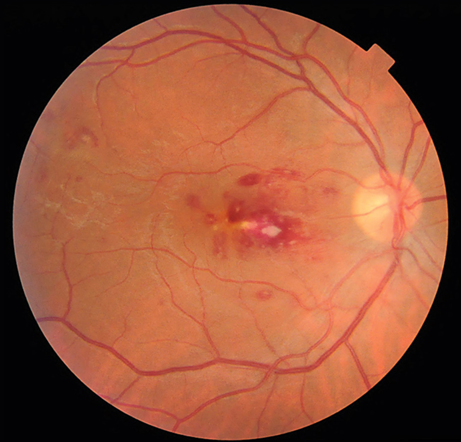
What is the most likely cause of these retinal changes?
What is the most likely diagnosis?
Diagnosis, Pathophysiology & OSCE Clinical Study Notes
Infective endocarditis — The presence of white-centered hemorrhages (Roth spots) should prompt the consideration of possible infective endocarditis. This patient's diagnosis was confirmed with echocardiography and blood cultures.
Infective endocarditis (IE) is a microbial infection of the endocardial surface of the heart, most commonly affecting the heart valves. It can also involve septal defects or the mural endocardium.
Pathophysiology
The process typically begins with damage to the endocardium, which can result from turbulent blood flow (e.g., due to valvular disease) or direct injury (e.g., from an indwelling catheter). This damaged surface promotes the formation of a non-bacterial thrombotic vegetation (platelet-fibrin thrombus). During periods of bacteremia, microorganisms can adhere to this thrombus and proliferate, forming an infected vegetation. Common causative organisms include Staphylococcus aureus (especially in IV drug users and healthcare-associated infections), Viridans group streptococci, and Enterococci.
Clinical Features
Presentation can be acute and fulminant or subacute and indolent. Key features include:
- Systemic: Fever, chills, night sweats, malaise, and weight loss are common.
- Cardiac: A new or changing heart murmur is a classic sign. Heart failure can develop due to valvular destruction.
- Peripheral Stigmata (Immunologic/Vascular): These classic signs are less common with early diagnosis but are important to recognize.
- Roth spots: White-centered retinal hemorrhages.
- Osler nodes: Painful, violaceous nodules on the fingers or toes.
- Janeway lesions: Painless, erythematous macules on the palms and soles.
- Splinter hemorrhages: Linear hemorrhages under the nails.
Management involves a prolonged course (typically 4-6 weeks) of high-dose intravenous antibiotics, tailored to the specific organism and its sensitivities. Surgical intervention may be required for complications such as severe heart failure, uncontrolled infection, large vegetations with high embolic risk, or valve destruction.
OSCE Quick Revision Q&A
What is the most likely diagnosis for "Roth Spots: An Ocular Red Flag"?
The most likely diagnosis is Infective endocarditis. The presence of white-centered hemorrhages (Roth spots) should prompt the consideration of possible infective endocarditis. This patient's diagnosis was confirmed with echocardiography and blood cultures.
Which medical OSCE exams test Infective endocarditis visual spot diagnosis?
Visual clinical sign recognition in Cardiology is regularly examined in Final MBBS OSCEs, UKMLA CPSA, PLAB 2, MRCP PACES, AMC Clinical, NAC OSCE, and USMLE/MD clerkship clinical exams.