A 62-year-old man presents with an incidental retroperitoneal mass on imaging. Discover the rare diagnosis behind the classic ‘hairy kidney’ sign.

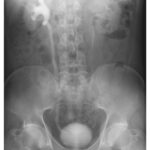
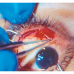
A 62-year-old man undergoing abdominal ultrasonography for the evaluation of gallstones was found to have a retroperitoneal mass. A physical examination and the results of routine laboratory studies were normal. A computed tomographic (CT) urogram was completed (left), and a subsequent positron-emission tomography–CT showed no hypermetabolic activity. He underwent stent placement in both ureters, and a core-biopsy of the perinephric soft tissue was obtained. Hematoxylin and eosin staining of the specimen is shown (right). What is the diagnosis?
What is the most likely diagnosis?
Erdheim–Chester disease, Hairy Kidney, Histiocytosis, Retroperitoneal Fibrosis, Urology