Painful Vision: A Neurological Clue
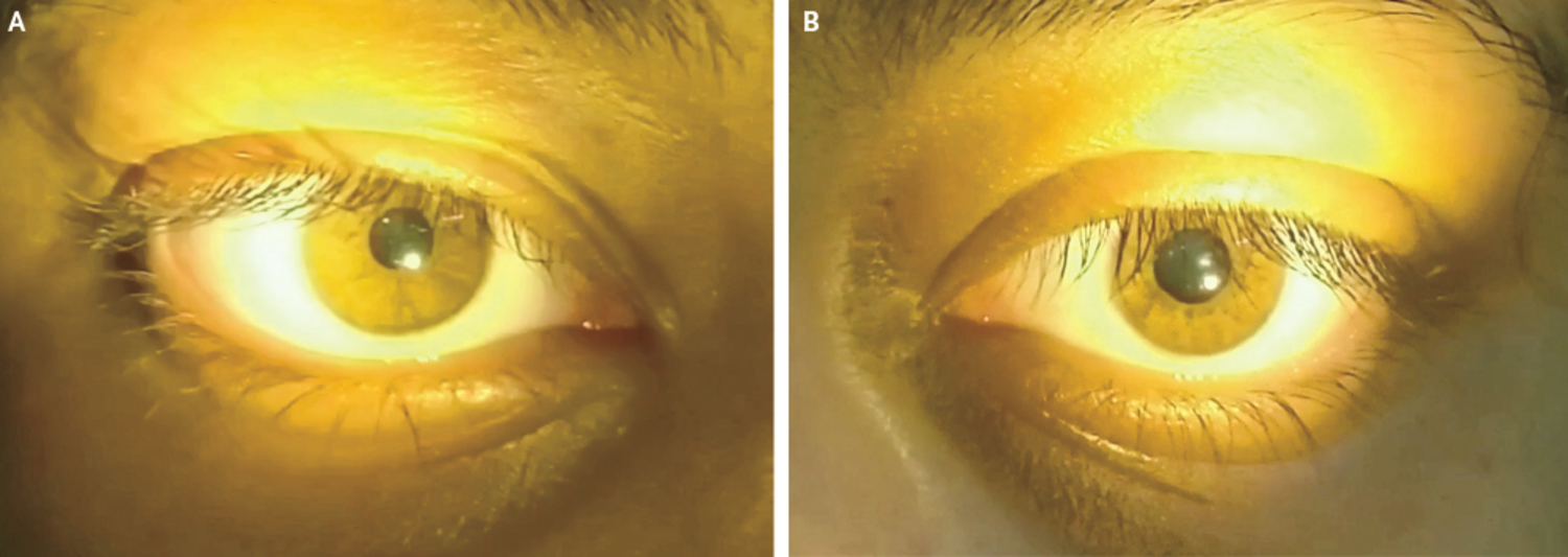
A 13-year-old girl presented with new onset blurry vision in the left eye associated with painful eye movements. Three weeks earlier, weakness and paresthesias had developed in her left leg that spontaneously resolved. When a light was swung from the normal right eye (Panel A) to the affected left eye, the left pupil dilated (Panel B) – a finding known as a relative afferent pupillary defect. Fundoscopic examination of both eyes was normal. Examination of the brain and whole spine by means of magnetic resonance imaging showed multiple, oblong hyperintense lesions on T2-weighted images. What is the most likely etiology?
What is the most likely diagnosis?
Diagnosis, Pathophysiology & OSCE Clinical Study Notes
Multiple sclerosis — The correct answer is multiple sclerosis. Serum tests for aquaporin-4 and myelin oligodendrocyte glycoprotein antibodies were negative. Oligoclonal bands were present in the cerebrospinal fluid. Symptoms resolved after the initiation of steroids followed by maintenance therapy with interferon beta-1a.
Multiple Sclerosis (MS) is a chronic, immune-mediated inflammatory disease of the central nervous system (CNS) characterized by demyelination, axonal damage, and neurodegeneration.
Pathophysiology
The primary pathology involves an autoimmune attack, led by T-cells, against the myelin sheath and the myelin-producing oligodendrocytes in the CNS. This leads to the formation of demyelinating plaques, disrupting nerve signal conduction and causing a wide range of neurological symptoms.
Clinical Features
The diagnosis requires demonstration of dissemination of lesions in time and space. Clinical presentations are highly variable and reflect the location of the CNS lesions.
- Optic Neuritis: Often a presenting feature, causing painful, subacute monocular vision loss and a Relative Afferent Pupillary Defect (RAPD).
- Transverse Myelitis: Sensory, motor, and Autonomic Dysfunction below the level of the spinal cord lesion.
- Brainstem Syndromes: Such as internuclear ophthalmoplegia (INO), diplopia, vertigo, and facial numbness.
- Sensory Symptoms: Paresthesias, numbness, and Lhermitte's sign (electric shock-like sensation down the spine on neck flexion).
- Motor Symptoms: Weakness, spasticity, and gait disturbance.
- Magnetic Resonance Imaging (MRI): The cornerstone of diagnosis. T2-weighted and FLAIR sequences show characteristic hyperintense lesions in periventricular, juxtacortical, infratentorial, and spinal cord locations. "Dawson's fingers" are classic periventricular lesions.
- Cerebrospinal Fluid (CSF): The presence of two or more unique oligoclonal bands in the CSF (not present in the serum) is found in >95% of patients and is highly supportive of the diagnosis.
- Exclusion of Mimics: Serology for NMO-IgG (aquaporin-4) and MOG-IgG is essential to rule out neuromyelitis optica spectrum disorder (NMOSD) and MOG antibody disease (MOGAD).
- Acute Relapses: Treated with high-dose corticosteroids (e.g., intravenous methylprednisolone).
- Disease-Modifying Therapies (DMTs): The mainstay of long-term treatment to reduce relapse rates and slow disability progression. Options range from injectable therapies (e.g., interferon beta, glatiramer acetate) to oral and infusion therapies.
- Symptomatic Treatment: Addresses specific symptoms like fatigue, spasticity, neuropathic pain, and bladder dysfunction.
OSCE Quick Revision Q&A
What is the most likely diagnosis for "Painful Vision: A Neurological Clue"?
The most likely diagnosis is Multiple sclerosis. The correct answer is multiple sclerosis. Serum tests for aquaporin-4 and myelin oligodendrocyte glycoprotein antibodies were negative. Oligoclonal bands were present in the cerebrospinal fluid. Symptoms resolved after the initiation of steroids followed by maintenance therapy with interferon beta-1a.
Which medical OSCE exams test Multiple sclerosis visual spot diagnosis?
Visual clinical sign recognition in Neurology is regularly examined in Final MBBS OSCEs, UKMLA CPSA, PLAB 2, MRCP PACES, AMC Clinical, NAC OSCE, and USMLE/MD clerkship clinical exams.