The Red Flag of a White Pupil
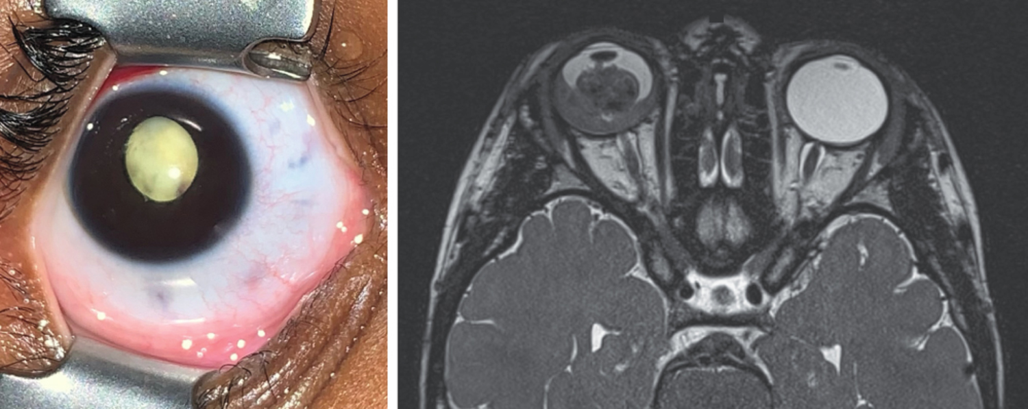
A 3-year-old girl was brought to the emergency department with a 2-month history of a white pupil and a 1-day history of redness and pain in the right eye. An eye examination showed leukocoria, as well as iris neovascularization and a white, nodular mass in the posterior chamber. The left eye was normal. B-scan ultrasonography showed calcification of the mass and vitreous seeding in the affected eye. Which of the following is the most likely diagnosis?
What is the most likely diagnosis?
Diagnosis, Pathophysiology & OSCE Clinical Study Notes
Retinoblastoma — A diagnosis of retinoblastoma was made. Magnetic resonance imaging of the head showed retinal and choroidal invasion without extraocular extension. Given the high-risk clinical features, the eye was enucleated the next day. Histopathological examination confirmed the diagnosis. Leukocoria requires urgent evaluation by an ophthalmologist in order to quickly identify life-threatening causes such as retinoblastoma.
Retinoblastoma is the most common primary intraocular malignancy in children, typically diagnosed before the age of 5. It arises from primitive retinal photoreceptor cells.
Pathophysiology
The tumor is caused by mutations in the RB1 tumor suppressor gene on chromosome 13q14. This follows a "two-hit" model:
- Heritable (40%): An inherited germline mutation (first hit) is present in all cells. A second, somatic mutation (second hit) in a retinal cell leads to tumor formation. These cases are often bilateral and multifocal.
- Sporadic (60%): Both mutations are somatic and occur in a single retinal cell. These cases are typically unilateral and unifocal.
- Leukocoria (approx. 60%): A white Pupillary Reflex, often noticed in flash photography ("cat's eye reflex"). This is the most common sign.
- Strabismus (approx. 20%): Misalignment of the eyes, which can be the first sign if the tumor affects the macula.
- Secondary Glaucoma: Presents as a red, painful eye with a fixed, dilated pupil, caused by tumor growth obstructing aqueous outflow.
- Fundoscopy: Dilated eye examination reveals a chalky-white retinal mass.
- Ultrasonography (B-scan): Highly useful for detecting intratumoral Calcification, a pathognomonic feature.
- Magnetic Resonance Imaging (MRI): The preferred imaging modality for assessing tumor size, optic nerve invasion, and extraocular extension, which is critical for staging.
- Enucleation (eye removal): Indicated for large tumors with no useful vision, neovascular glaucoma, or invasion of the optic nerve.
- Globe-Sparing Therapies: For smaller tumors, this may include systemic or intra-arterial chemotherapy, laser photocoagulation, cryotherapy, or plaque radiotherapy.
OSCE Quick Revision Q&A
What is the most likely diagnosis for "The Red Flag of a White Pupil"?
The most likely diagnosis is Retinoblastoma. A diagnosis of retinoblastoma was made. Magnetic resonance imaging of the head showed retinal and choroidal invasion without extraocular extension. Given the high-risk clinical features, the eye was enucleated the next day. Histopathological examination confirmed the diagnosis. Leukocoria requires urgent evaluation by an ophthalmologist in order to quickly identify life-threatening causes such as retinoblastoma.
Which medical OSCE exams test Retinoblastoma visual spot diagnosis?
Visual clinical sign recognition in Ophthalmology is regularly examined in Final MBBS OSCEs, UKMLA CPSA, PLAB 2, MRCP PACES, AMC Clinical, NAC OSCE, and USMLE/MD clerkship clinical exams.